Clinical Toxicology
Acquired Methemoglobinemia: Causes, Diagnosis, and Treatment
Amirhosein Shabrang
Post on 03 Sept 2026 · Updated 02 Oct 2026 · 20 min read
Amirhosein Shabrang
Post on 03 Sept 2026 · Updated 02 Oct 2026 · 20 min read
 Medical Toxicologymedicaltoxic.com
https://medicaltoxic.com/blogs/acquired-methemoglobinemia
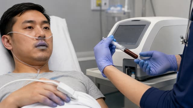
A patient is cyanotic.
The pulse oximeter remains unexpectedly low despite supplemental oxygen.
An arterial blood gas may show a surprisingly preserved partial pressure of oxygen.
The lungs may not explain the severity of the apparent hypoxemia.
When those findings do not fit together, clinicians should think beyond ordinary pulmonary oxygenation failure.
One important possibility is acquired methemoglobinemia.
Methemoglobinemia occurs when the iron in hemoglobin is oxidized from its normal ferrous state, Fe²⁺, to the ferric state, Fe³⁺. Ferric heme cannot bind oxygen normally, and the remaining functional hemoglobin holds oxygen more tightly. The result is impaired oxygen transport and tissue delivery—a form of functional anemia that can become life-threatening even when oxygen is reaching the lungs. [1, 2]
Acquired disease is usually triggered by an oxidizing medication, chemical, or toxin.
Recognizing it matters because standard pulse oximetry and ordinary blood-gas interpretation can be misleading, while the treatment—methylene blue—has important limitations and toxicities of its own.
Acquired methemoglobinemia results from oxidation of hemoglobin iron from Fe²⁺ to Fe³⁺, reducing effective oxygen carriage and delivery.
Important causes include dapsone, benzocaine and prilocaine, nitrates and nitrites, aniline-related chemicals, propanil, recreational alkyl nitrites, and selected other oxidant exposures.
Persistent cyanosis or an unexpectedly low SpO₂ despite supplemental oxygen should raise suspicion, particularly after a compatible exposure.
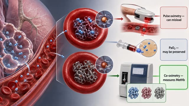
PaO₂ may remain normal or high because it measures oxygen dissolved in plasma, not whether hemoglobin can carry oxygen normally.
A saturation gap is a useful diagnostic clue but is not required and should not be treated as a universal hallmark.
Co-oximetry is the diagnostic standard for measuring methemoglobin in a clinically suspected case.
Methylene blue is the principal antidotal treatment for clinically significant acquired methemoglobinemia, but treatment decisions should reflect symptoms, physiology, comorbidities, exposure, and MetHb concentration—not one universal percentage.
Current U.S. labeling recommends methylene blue 1 mg/kg IV over 5–30 minutes, with carefully defined criteria for one repeat dose.
Current U.S. labeling lists known G6PD deficiency as a contraindication because methylene blue may be ineffective and may cause severe hemolysis.
Methylene blue is a potent MAO-A inhibitor and can precipitate serotonin toxicity in patients exposed to relevant serotonergic drugs.
Dapsone, aniline-related compounds, and selected agrochemical poisonings can produce prolonged or rebound methemoglobinemia.
Exchange transfusion and hyperbaric oxygen are rescue options in selected severe cases when methylene blue fails; ascorbic acid is slower and should not be presented as an equivalent emergency substitute.
Normal hemoglobin depends on iron remaining predominantly in the ferrous Fe²⁺ state so oxygen can bind reversibly.
Oxidant stress converts part of that iron to ferric Fe³⁺.
The affected heme groups can no longer bind oxygen effectively. At the same time, oxygen binds more tightly to the remaining functional sites, making unloading to tissues more difficult. [2, 3]
Red blood cells constantly generate small amounts of methemoglobin during normal physiology. Endogenous reducing systems—particularly cytochrome b5 reductase—normally convert it back to functional hemoglobin.
Acquired methemoglobinemia develops when oxidant exposure overwhelms those protective systems.
Congenital methemoglobinemia can result from cytochrome b5 reductase deficiency or hemoglobin M variants.
That distinction matters diagnostically, but congenital disease should not dominate an acute toxicology article.
In a patient with new cyanosis after a medication, chemical, recreational, occupational, or agricultural exposure, an acquired oxidant process is the immediate clinical concern.
A lifelong history of cyanosis, family history, or persistent unexplained MetHb after the exposure has resolved should prompt evaluation for inherited disease. [3]
There is no clinical advantage to memorizing an enormous list of every agent ever mentioned in a case report.
A better approach is to recognize the exposure groups with the strongest and most useful human evidence.
Dapsone is one of the most important causes because toxicity can be both substantial and prolonged.
Its oxidizing metabolites can persist after the parent drug has been absorbed, creating recurrent MetHb elevation after an initially successful response to treatment.
A 2026 ten-year retrospective cohort from a Thai poison center included 169 patients with acquired methemoglobinemia. Dapsone was the most common identified agent in that cohort, followed closely by the herbicide propanil. [4]
Those proportions are specific to that population and should not be interpreted as global incidence.
Benzocaine is a well-established cause of acquired methemoglobinemia, particularly after topical or oropharyngeal use.
Prilocaine can also produce clinically significant methemoglobinemia.
Lidocaine is much more strongly associated with local-anesthetic systemic neurotoxicity and cardiotoxicity, but methemoglobinemia can occur in relevant circumstances. MedicalToxic addresses those distinctions in Viscous Lidocaine Poisoning: An Evidence-Based Guideline.
Topical anesthetics are also discussed within the broader exposure framework of Personal Care Cosmetics and Topical Products Management.
Nitrate and nitrite exposures are classic causes of oxidant stress.
Potential sources include medications, chemicals, contaminated products, and intentional ingestion.
Recreational alkyl nitrites—often called “poppers”—have also become clinically relevant. In a 2025 retrospective study of methylene-blue-treated cases reported to the New York City Poison Center, volatile nitrites were the largest exposure group, followed by local anesthetics and dapsone. [5]
Because that study included only cases in which methylene blue was administered, its exposure distribution should not be interpreted as population incidence.
Aniline and related aromatic amines are important occupational and toxicologic oxidants.
These exposures deserve particular attention because methemoglobinemia may persist or rebound after apparently successful treatment. Current methylene-blue labeling specifically warns about recurrence with aryl amines such as aniline. [6]
Propanil is a well-documented agricultural cause of methemoglobinemia and represented a substantial proportion of cases in the 2026 Thai cohort. [4]
The differential is now broader.
A 2026 five-patient Clinical Toxicology case series described methemoglobinemia after several uncommon agrochemical products, including propanil, 2,4-D ethyl ester, indoxacarb, and two exposures involving a seaweed-extract biostimulant. All five patients had oxygen-unresponsive hypoxia with a saturation gap, and the clinical courses included rebound methemoglobinemia, intravascular hemolysis, and acute kidney injury. [7]
Those five cases are signals from a small case series, not evidence that entire pesticide or agrochemical classes share the same risk.
For the research-specific discussion, see New Case Series Links Uncommon Agrochemicals to Acquired Methemoglobinemia.
The clinical spectrum ranges from an abnormal color and pulse-oximetry reading to critical impairment of oxygen delivery.
Possible manifestations include:
cyanosis;
headache;
fatigue;
dizziness;
dyspnea;
tachycardia;
confusion;
chest discomfort;
syncope;
seizures;
dysrhythmias;
metabolic acidosis;
coma;
cardiovascular collapse.
Severity cannot be predicted by MetHb percentage alone.
A patient with anemia, coronary disease, pulmonary disease, reduced cardiac output, or another impairment of oxygen delivery may develop clinically important symptoms at a lower concentration than a healthy patient. Rate of MetHb formation also matters. [2, 3]
Cyanosis is the classic finding.
But relying on it as a requirement can delay diagnosis.
A systematic review of 87 published acquired-methemoglobinemia cases found cyanosis in 82% and pulse-oximetry saturation below 90% in 60%. [8]
Those figures come from published case reports and are heavily affected by selection and publication bias. They are not estimates of sensitivity or real-world incidence.
The useful conclusion is simply:
classic signs are common, but they are not universal.
Dark or “chocolate-brown” blood is a classic visual clue.
When present, it can reinforce suspicion.
But color is subjective and can be influenced by lighting, hemoglobin concentration, and other dyshemoglobins. It should not replace direct laboratory measurement. [2]
Standard pulse oximeters estimate hemoglobin saturation using light at a limited number of wavelengths.
They are designed primarily to distinguish oxyhemoglobin from deoxyhemoglobin.
Methemoglobin absorbs light differently and interferes with that calculation. As MetHb rises, conventional pulse oximetry becomes increasingly unreliable rather than providing an accurate measure of tissue oxygen delivery. [2]
This explains why simply adding more oxygen may not make the SpO₂ behave as expected.
Supplemental oxygen can still be clinically useful.
It increases dissolved oxygen and maximizes oxygen availability to the hemoglobin that remains functional.
It just does not chemically reduce Fe³⁺ back to Fe²⁺.
PaO₂ measures oxygen dissolved in plasma.
It does not directly measure whether hemoglobin is capable of transporting that oxygen normally.
A patient with significant methemoglobinemia may therefore have a preserved—or, during oxygen administration, very high—PaO₂ while oxygen delivery by hemoglobin remains impaired. [1, 2]
That is one of the most important diagnostic traps.
A normal PaO₂ does not exclude methemoglobinemia.
A saturation gap describes discordance between oxygen saturation measurements obtained by different methods—commonly an unexpectedly low pulse-oximetry reading compared with a saturation calculated from arterial PaO₂.
The reason matters.
Many routine blood-gas analyzers calculate oxygen saturation from PaO₂ using assumptions based on normal hemoglobin.
That calculated value may therefore appear reassuring even when a clinically important dyshemoglobin is present.
This mismatch can be a powerful clue.
But it is not a diagnostic requirement.
Recent clinical review and the systematic review of published cases both emphasize that the expected saturation-gap pattern is not consistently present in every patient. [2, 8]
Therefore:
absence of a textbook saturation gap should not be used to rule out methemoglobinemia when the exposure and clinical picture remain convincing.
When clinically significant methemoglobinemia is suspected, MetHb should be measured by co-oximetry.
Co-oximeters use multiple wavelengths of light to distinguish different hemoglobin species rather than assuming that all hemoglobin exists only as oxyhemoglobin or deoxyhemoglobin.
This allows direct estimation of:
oxyhemoglobin;
deoxyhemoglobin;
methemoglobin;
carboxyhemoglobin;
with capabilities varying by analyzer.
Contemporary review identifies co-oximetry as the diagnostic standard when methemoglobinemia is clinically suspected. [2]
Even a sophisticated laboratory measurement should be interpreted in context.
Sulfhemoglobin can also produce cyanosis and dark blood. Its absorption spectrum overlaps with methemoglobin sufficiently that older co-oximeters may misclassify sulfhemoglobin as MetHb. [2]
If the reported MetHb level, exposure history, clinical picture, and treatment response do not fit together, consultation with the laboratory and more specific testing may be necessary.
That is a more useful rule than treating every co-oximeter number as infallible.
The bedside reasoning can be organized around four questions.
Look for:
unexplained cyanosis;
persistently abnormal SpO₂;
weak apparent response of the SpO₂ to oxygen;
a PaO₂ that seems disproportionately reassuring;
symptoms suggesting impaired oxygen delivery.
Ask specifically about:
dapsone;
topical or local anesthetics;
dental or endoscopic procedures;
nitrates and nitrites;
recreational alkyl nitrites;
pesticides and herbicides;
occupational chemicals;
dyes or aromatic amines;
intentional ingestion;
unfamiliar medications or consumer products.
Obtain co-oximetry when clinically significant methemoglobinemia is suspected.
Important alternatives include:
conventional pulmonary hypoxemia;
carbon monoxide poisoning;
sulfhemoglobinemia;
low-perfusion or motion-related pulse-oximeter artifact;
congenital dyshemoglobinemia.
The diagnosis is built from exposure + physiology + direct measurement, not from one monitor.
Stop the oxidant exposure whenever possible.
Support airway, breathing, circulation, and tissue oxygen delivery.
Provide supplemental oxygen when clinically indicated.
Then assess whether the patient requires antidotal therapy.
Methylene blue should not be withheld from a clinically deteriorating patient simply because an arbitrary numerical threshold has not been crossed.
Likewise, an isolated MetHb percentage should not automatically trigger treatment without considering symptoms and clinical reserve.
The 2025 American Heart Association guideline gives a Class 1 recommendation for methylene blue in adults and children with life-threatening methemoglobinemia. [1]
Broader hematology guidance similarly integrates MetHb concentration with symptoms, cause, age, anemia, and cardiopulmonary status. [3]
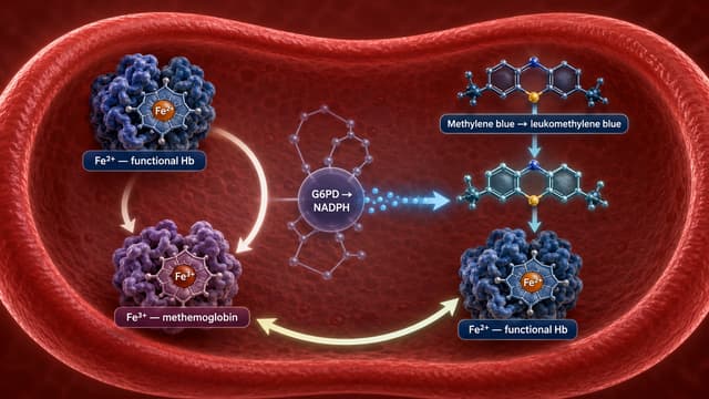
Methylene blue provides an alternative pathway for reducing methemoglobin.
Within red blood cells, NADPH generated through the pentose-phosphate pathway helps convert methylene blue to its reduced form, which then promotes conversion of ferric Fe³⁺ methemoglobin back toward functional ferrous Fe²⁺ hemoglobin. [3]
This mechanism explains both its therapeutic effect and one of its most important limitations: dependence on adequate NADPH production.
Because dosing is high consequence, this section uses the current U.S. prescribing information, revised January 2026.
For adults and children with acquired methemoglobinemia, current U.S. labeling recommends:
1 mg/kg intravenously over 5–30 minutes. [6]
If the MetHb level remains above 30% or clinical signs and symptoms persist, one additional 1 mg/kg dose may be given one hour after the first dose. If methemoglobinemia has not resolved after two doses, the label recommends moving to alternative interventions rather than continuing routine repeated dosing. [6]
An important distinction:
the >30% value in the U.S. label is a criterion for repeat dosing. It is not a universal threshold that must be reached before initial treatment.
The current U.S. label recommends only a single 1 mg/kg dose in patients with moderate or severe renal impairment, defined there as eGFR 15–59 mL/min/1.73 m².
If significant MetHb elevation or symptoms persist after that dose, alternative treatment should be considered rather than routine redosing. [6]
This is a useful example of why antidote dosing should be checked against the current product information rather than copied from an older toxicology summary.
Outside the United States, clinicians should use the current local product information and poison-center or medical-toxicology protocol.
Methylene blue has long-standing clinical use, but randomized outcome trials are lacking.
The AHA recommendation is supported by observational evidence and accumulated clinical experience rather than randomized trials. [1]
A useful contemporary dataset comes from a 2025 retrospective study of cases reported to the New York City Poison Center over 24 years.
The study included 185 patients who received methylene blue for methemoglobinemia. Clinical improvement was reported in most cases, and the median administered dose was 1 mg/kg. Multiple doses were used in a minority of patients. [5]
This strengthens real-world evidence that methylene blue is generally effective.
But it is still a retrospective, treatment-selected poison-center cohort—not a randomized comparison and not evidence that every patient with an elevated MetHb level requires treatment.
Glucose-6-phosphate dehydrogenase helps generate the NADPH required for methylene blue's reduction pathway.
In G6PD deficiency, two problems arise:
methylene blue may be less effective because conversion to its active reduced form is impaired;
oxidative stress from methylene blue can contribute to clinically important hemolysis.
Current U.S. methylene-blue labeling lists known G6PD deficiency as a contraindication because of the risk of hemolytic anemia. It also warns that treatment may be ineffective. [6]
The AHA similarly notes both potential treatment failure and hemolysis in G6PD-deficient patients. [1]
That should not be converted into vague reassurance or into a statement about every possible international product label.
The practical message is:
known or strongly suspected G6PD deficiency materially changes antidote planning.
In a patient with severe methemoglobinemia and this complication, involve medical toxicology and hematology early and consider the available rescue strategies.
Methylene blue is not merely a redox drug.
It is also a potent reversible inhibitor of monoamine oxidase A (MAO-A). Experimental pharmacology demonstrated clinically relevant MAO-A inhibition, providing the mechanistic basis for serotonin toxicity when methylene blue is combined with serotonergic drugs. [9]
Current U.S. labeling carries a boxed warning for serotonin syndrome with concomitant serotonergic medications and opioids and also identifies dextromethorphan as a relevant risk. [6]
Medication reconciliation therefore matters before methylene blue whenever clinical circumstances permit.
Look specifically for:
SSRIs;
SNRIs;
MAO inhibitors;
serotonergic opioids;
dextromethorphan;
other clinically meaningful serotonergic combinations.
This becomes a difficult risk-benefit problem when severe methemoglobinemia itself is immediately life-threatening. Such cases warrant specialist toxicology input and consideration of alternative strategies rather than treating the interaction as a routine prescribing issue.
For recognition and management of serotonin toxicity, see Serotonin Syndrome (Serotonin Toxicity): Symptoms, Diagnosis, Drug Interactions, and Treatment.
A rapid fall in MetHb after methylene blue does not always mean the poisoning is finished.
Current U.S. labeling specifically warns that methemoglobinemia may persist or rebound after apparently successful treatment when the oxidant is an aryl amine such as aniline or a sulfonamide drug such as dapsone. [6]
Rebound also appeared in the 2026 agrochemical case series. [7]
Possible mechanisms include:
continued absorption;
delayed formation of oxidizing metabolites;
prolonged drug elimination;
persistent exposure;
or a product containing more than one relevant component.
Serial co-oximetry is therefore particularly important when the causative agent has prolonged toxicokinetics.
Methemoglobinemia and hemolysis can occur together, but they should not be treated as the same process.
The underlying oxidant can injure red cells.
G6PD deficiency can increase susceptibility.
Methylene blue itself can cause hemolysis.
Current U.S. labeling notes that methylene-blue-associated anemia may be delayed by one or more days and can be severe enough to require red-cell transfusion. [6]
In the 2026 agrochemical case series, selected patients developed intravascular hemolysis and acute kidney injury. [7]
Those complications should not be generalized to every acquired-methemoglobinemia patient.
They do mean that when an oxidant poisoning has hemolytic potential, follow-up may need to include hemoglobin, bilirubin, haptoglobin, renal function, urine findings, and other testing guided by the clinical course.
There is no single rescue strategy backed by the same level of clinical experience as methylene blue.
AHA 2025 states that exchange transfusion may be reasonable for adults and children with life-threatening methemoglobinemia that does not respond to methylene blue. [1]
Evidence is mainly case-based.
This is a rescue therapy, not routine first-line treatment.
Hyperbaric oxygen may also be reasonable for life-threatening disease that is unresponsive to methylene blue. [1]
Its limitations are important:
evidence is limited;
reduction of MetHb may take hours;
access may be restricted;
transfer may be inappropriate for an unstable patient.
Ascorbic acid can reduce methemoglobin through a slower nonenzymatic process.
AHA 2025 states that it may be reasonable in life-threatening methemoglobinemia when methylene blue is contraindicated or unavailable. [1]
Its action is too slow to present it as an equivalent substitute for methylene blue in a rapidly deteriorating patient.
AHA 2025 specifically recommends against N-acetylcysteine as treatment for life-threatening methemoglobinemia because available evidence does not support clinical benefit. [1]
The responsible oxidant should determine much of the monitoring strategy.
Important components may include:
serial clinical examination;
oxygenation and cardiovascular monitoring in significant cases;
repeat co-oximetry;
monitoring for recurrent MetHb elevation;
assessment for hemolysis when relevant;
renal function when hemolysis, pigment injury, or the specific poison makes AKI plausible;
acid-base status in severe illness.
There is no universal observation period.
A short-lived iatrogenic local-anesthetic exposure is not the same toxicokinetic problem as dapsone or propanil poisoning.
Higher-acuity or intensive monitoring is appropriate when there is:
significant neurological or cardiovascular toxicity;
severe impairment of oxygen delivery;
recurrent MetHb elevation;
hemodynamic instability;
respiratory support requirement;
need for repeated or rescue treatment;
substantial hemolysis;
acute kidney injury or other organ dysfunction.
Disposition should be based on clinical recovery, serial MetHb behavior, the responsible agent, and the likelihood of delayed or recurrent toxicity rather than a fixed number of hours.
Supplemental oxygen does not chemically reduce methemoglobin.
The SpO₂ may remain abnormal even while oxygen availability to functional hemoglobin improves.
PaO₂ measures dissolved oxygen.
It does not tell you whether hemoglobin is functioning normally.
The saturation gap is supportive, not mandatory.
Its absence does not overrule a compatible exposure and co-oximetry result. [2, 8]
Conventional pulse oximetry does not reliably quantify methemoglobin.
Use co-oximetry.
Older analyzers may confuse sulfhemoglobin with methemoglobin.
If the result does not fit the history or treatment response, investigate the discrepancy. [2]
Anemia, heart disease, pulmonary disease, rate of MetHb formation, ongoing exposure, and symptoms all modify clinical risk.
Methylene blue is itself a strongly colored compound.
Current labeling warns that it can transiently cause underestimation of oxygen saturation by pulse oximetry after administration. [6]
Do not misinterpret a post-treatment monitor artifact as proof that the patient has worsened.
Methylene blue can create a new toxicologic problem in a patient taking serotonergic medications.
Medication reconciliation belongs in the antidote decision.
Dapsone, aniline-related compounds, and selected agrochemical exposures can produce recurrent methemoglobinemia.
Low SpO₂ plus a surprisingly adequate PaO₂ should trigger consideration of dyshemoglobinemia.
Co-oximetry answers a different question from an ordinary blood gas.
The saturation gap is a clue, not a diagnostic requirement.
Do not let one MetHb percentage replace the patient's physiology.
The causative poison matters after the MetHb falls. Some agents can produce rebound, hemolysis, or other systemic toxicity.
Methylene blue has its own toxicology. G6PD status, renal function, serotonergic drugs, and repeat dosing all matter.
A successful antidote response does not eliminate the need for monitoring.
PaO₂ measures oxygen dissolved in plasma. Methemoglobinemia primarily interferes with hemoglobin's ability to transport and release oxygen. A reassuring PaO₂ therefore does not prove that total oxygen delivery is adequate.
It is an unexpected discrepancy between oxygen-saturation measurements obtained by different methods, commonly a low pulse-oximetry reading compared with a saturation calculated from arterial PaO₂. It supports suspicion for dyshemoglobinemia but is not present in every case.
Measurement of MetHb with co-oximetry is the diagnostic standard when clinically significant methemoglobinemia is suspected. [2]
There is no single universal treatment threshold for every patient. Symptoms, cardiovascular and pulmonary reserve, anemia, rate of progression, ongoing exposure, and the measured MetHb concentration should be considered together.
For life-threatening methemoglobinemia, AHA 2025 recommends methylene blue. [1]
Current U.S. labeling recommends 1 mg/kg IV over 5–30 minutes. If MetHb remains above 30% or clinical symptoms persist, one additional 1 mg/kg dose may be given one hour later. Patients with moderate or severe renal impairment have a different current labeled redosing strategy. [6]
Current U.S. labeling lists known G6PD deficiency as a contraindication because methylene blue may be ineffective and can cause severe hemolytic anemia. Severe methemoglobinemia in this setting requires individualized toxicology/hematology management and consideration of rescue alternatives. [6]
Yes. Methylene blue inhibits MAO-A, and current U.S. labeling carries a boxed warning regarding serious or fatal serotonin syndrome with relevant serotonergic co-medications. [6, 9]
Acquired methemoglobinemia is not simply a low oxygen-saturation number.
It is a disorder of functional oxygen transport.
The clue is often a mismatch:
the patient may be cyanotic;
the pulse oximeter may remain unexpectedly low;
supplemental oxygen may not make the monitor behave normally;
and the PaO₂ may look much more reassuring than expected.
When those findings do not make physiologic sense together, clinicians should consider dyshemoglobinemia and measure the hemoglobin species directly.
Co-oximetry is the diagnostic pivot.
Treatment then depends on the patient, the responsible oxidant, and the severity of impaired oxygen delivery—not on a single universal MetHb threshold.
Methylene blue remains the principal antidotal treatment for clinically important acquired methemoglobinemia, but it must be used with the same toxicologic discipline applied to the poison itself. Current dosing, renal function, G6PD deficiency, serotonergic medications, hemolysis, and rebound all matter.
And when methylene blue cannot be used or does not work, exchange transfusion, hyperbaric oxygen, or slower alternative reduction strategies may have roles in carefully selected severe cases.
The bedside lesson is straightforward:
when the oxygen numbers do not fit the patient, think beyond the lungs—and measure the hemoglobin species directly.
© All copyright of this material is absolute to Medical toxicology
As the Growth & Content Lead at MedicalToxic.com, I work on content strategy, development, and digital growth. My focus is on making MedicalToxic’s content clear, practical, and accessible, while helping expand the platform’s reach and strengthen its position as a trusted resource in clinical toxicology.
Keep reading
 Clinical Toxicology
Amirhosein Shabrang
 Clinical Toxicology
Amirhosein Shabrang
 Clinical Toxicology
Amirhosein Shabrang
 Clinical Toxicology
Amirhosein Shabrang