Toxic Agents and Antidotes
Diquat Poisoning: Neurotoxicity, Acute Kidney Injury, Prognosis, and Treatment
Amirhosein Shabrang
Post on 08 Oct 2026 · 18 min read
Amirhosein Shabrang
Post on 08 Oct 2026 · 18 min read
 Medical Toxicologymedicaltoxic.com
https://medicaltoxic.com/blogs/diquat-poisoning-neurotoxicity-acute-kidney-injury-prognosis
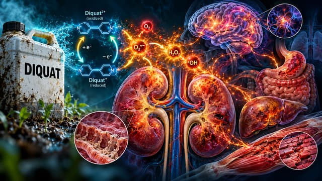
Diquat is a bipyridyl herbicide capable of causing severe systemic poisoning after substantial exposure, particularly after ingestion of concentrated liquid formulations.
Although Diquat is structurally related to paraquat, the two poisons should not be treated as interchangeable. Diquat undergoes redox cycling and generates reactive oxygen species, but its human toxicity is characterized particularly by gastrointestinal injury, acute kidney injury, hepatic and skeletal-muscle injury, neurotoxicity, shock, and multiorgan dysfunction. The delayed progressive pulmonary fibrosis that defines severe paraquat poisoning is not the dominant syndrome of Diquat toxicity. [1]
Recent human evidence has significantly improved understanding of the syndrome. A 2025 multicenter prospective study of 204 patients developed severity indices for triage and prognosis, while a 2026 cohort of 129 confirmed cases better characterized toxic encephalopathy and its imaging patterns. [2,3]
At the same time, mechanistic research is expanding rapidly. New studies implicate lipid peroxidation, mitochondrial dysfunction, ferroptosis-related pathways, and disturbances in serum lipid metabolism. These findings are important for understanding Diquat injury, but none currently establishes a new antidote or validated biomarker-based treatment pathway.
Diquat is a potent redox-cycling bipyridyl herbicide that generates reactive oxygen species and can produce severe multiorgan injury. [1]
The kidney is a major target organ. Acute tubular injury, oliguria, electrolyte disturbances, and kidney failure can develop after significant poisoning. [1,4]
Diquat and paraquat share chemical features but do not have identical clinical toxicity; paraquat-specific management assumptions should not automatically be transferred to Diquat.
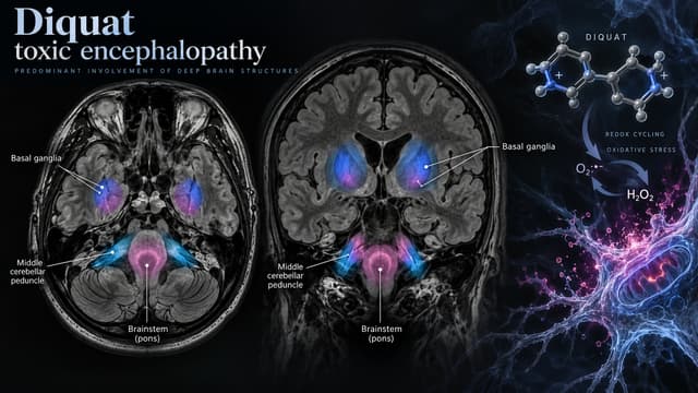
Severe neurotoxicity can include altered consciousness, seizures, coma, and toxic encephalopathy. A 2026 cohort identified frequent involvement of the brainstem, bilateral cerebellar peduncles, and basal ganglia in affected patients. [3]
Plasma Diquat concentration may provide prognostic information, but its meaning depends strongly on the time between ingestion and blood sampling. [5]
Two 2025 severity indices—SIDP-T and SIDP-P—show promising external validation for mortality risk assessment but should not yet be treated as universal triage standards. [2]
There is no established specific antidote for Diquat poisoning. Management remains centered on early assessment, selected decontamination, serial monitoring, and aggressive supportive care. [1,6]
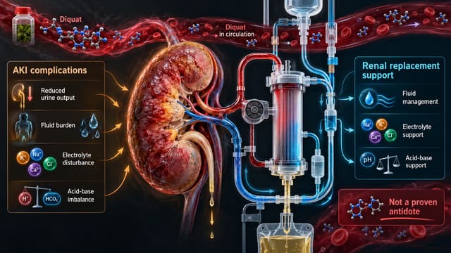
Renal replacement therapy can be lifesaving for complications of acute kidney injury, but it should not be presented as a proven Diquat antidote or reliable survival-improving detoxification strategy. [6]
Ferroptosis and lipid peroxidation are important research directions, but ferrostatin-1 has only been studied in Diquat-exposed cells—not as a treatment in poisoned patients.
Newly reported lipidomic and metabolomic markers remain exploratory and are not validated diagnostic or prognostic tests for routine clinical use.
Diquat, chemically 1,1′-ethylene-2,2′-bipyridinium, is a nonselective contact herbicide used for control of terrestrial and aquatic vegetation and as a crop desiccant.
Human poisoning has most commonly followed intentional ingestion of concentrated liquid formulations. Dermal, ocular, and inhalational exposures are also possible, but severe systemic poisoning is most strongly associated with substantial ingestion. [1]
The exact formulation matters. Commercial herbicide products may contain different concentrations, solvents, surfactants, or additional active ingredients, so clinicians should obtain the container, label, product name, and formulation whenever possible.
Diquat and paraquat are both bipyridyl herbicides and both undergo intracellular redox cycling.
That similarity has historically led to clinical extrapolation between the two poisons.
But their toxicologic profiles differ.
Feature | Diquat | Paraquat |
|---|---|---|
Core mechanism | Redox cycling / oxidative stress | Redox cycling / oxidative stress |
Major human targets | GI tract, kidney, nervous system, liver, muscle | Lung, kidney, GI tract, multiple organs |
AKI | Prominent | Common |
Neurotoxicity | Important in severe poisoning | Less defining |
Delayed pulmonary fibrosis | Not the defining syndrome | Characteristic severe complication |
Specific antidote | None established | None established |
Management | Supportive, Diquat-specific evidence required | Separate paraquat evidence base |
The practical point is simple:
Diquat is not merely “paraquat with less lung toxicity.”
Its renal and neurologic profile deserves independent assessment, and therapies used in paraquat poisoning should not automatically be assumed effective for Diquat. [1]
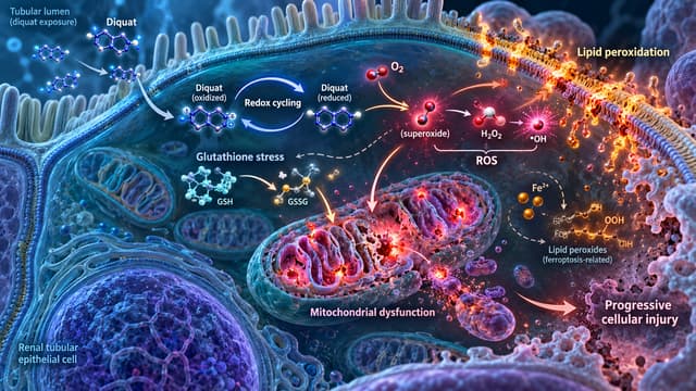
The best-established mechanism is redox cycling.
Diquat accepts an electron to form an unstable radical. That radical transfers the electron to molecular oxygen, generating superoxide while regenerating the original Diquat molecule. The toxin can therefore repeatedly cycle through this process while cellular reducing equivalents remain available. [1]
The downstream consequences include:
excessive reactive oxygen species;
glutathione and antioxidant-system stress;
lipid peroxidation;
mitochondrial injury;
membrane dysfunction;
inflammatory signaling;
cellular necrosis and other forms of cell death.
This mechanism helps explain why Diquat injury can affect several organ systems simultaneously rather than behaving like a toxin directed at one receptor.
After absorption, Diquat distributes systemically and is eliminated predominantly through the kidneys.
Renal handling makes the kidney both an important elimination pathway and a major site of toxicity. Acute renal tubular injury can reduce toxin elimination while simultaneously producing fluid, electrolyte, and acid-base complications. [1,4]
Human renal biopsy evidence remains limited, but a 2023 report of three patients with acute Diquat poisoning documented acute kidney injury in all three; biopsies in two showed acute tubular necrosis with interstitial edema and inflammatory-cell infiltration. [4]
For clinicians, this means an initially normal creatinine concentration does not rule out subsequent severe renal injury.
Serial measurements matter.
The clinical course varies according to formulation, dose, absorption, treatment delay, and patient factors.
Early gastrointestinal manifestations may include:
burning pain involving the mouth, throat, chest, or abdomen;
nausea;
repeated vomiting;
diarrhea;
gastrointestinal bleeding;
ileus in severe poisoning.
Direct mucosal injury and substantial fluid loss can contribute to dehydration and hemodynamic instability.
Severe gastrointestinal injury also complicates decontamination decisions because vomiting, impaired consciousness, and damaged mucosa increase procedural risk.
AKI is one of the central manifestations of severe Diquat poisoning.
Potential clinical findings include:
rising creatinine;
oliguria or anuria;
electrolyte abnormalities;
metabolic derangement;
fluid accumulation;
progression to kidney failure requiring renal replacement therapy.
Mechanisms likely include direct oxidative tubular injury, hemodynamic compromise, dehydration, mitochondrial dysfunction, and additional stress from rhabdomyolysis. [1,4]
Serum aminotransferases may rise during severe poisoning.
Skeletal-muscle injury and rhabdomyolysis have also been reported, with increases in creatine kinase and myoglobin. Muscle breakdown may further aggravate renal dysfunction and complicate interpretation of aminotransferase abnormalities.
Serial CK, renal function, electrolytes, urine output, and acid-base assessment can therefore be important in significant systemic poisoning.
Diquat neurotoxicity is increasingly recognized as a major feature of severe poisoning rather than a rare curiosity.
Manifestations may include:
dizziness;
drowsiness;
agitation or disorientation;
altered mental status;
seizures;
coma;
focal or diffuse neurologic abnormalities.
The strongest recent dataset is a 2026 retrospective cohort of 129 analytically confirmed acute Diquat poisonings. Forty-five patients developed toxic encephalopathy and 34 patients died; 27 of those who died had developed encephalopathy. [3]
These proportions describe one hospital cohort and should not be interpreted as the population incidence of encephalopathy or death after all Diquat exposures.
Among patients with Diquat toxic encephalopathy, reported abnormalities particularly involved:
the brainstem;
bilateral cerebellar peduncles;
basal ganglia. [3]
This pattern may provide a useful diagnostic clue when a patient deteriorates neurologically after confirmed or suspected Diquat poisoning.
Imaging should not, however, be treated as a mandatory screening test for every exposure. It becomes especially relevant when altered consciousness, seizures, focal findings, or otherwise unexplained neurologic deterioration occurs.
In the 129-patient cohort, toxic encephalopathy was strongly associated with death, with an odds ratio of 16.5. [3]
That does not prove that encephalopathy itself causes death.
It is more appropriately interpreted as a marker of severe systemic poisoning within that cohort.
Diagnosis begins with the exposure history and clinical trajectory.
Important questions include:
What exact herbicide was involved?
Was Diquat the only active ingredient?
What concentration was on the product label?
Was exposure oral, dermal, ocular, or inhalational?
When did exposure occur?
Was the estimated amount large or uncertain?
Was the exposure intentional?
Were any medications, pesticides, or other chemicals co-ingested?
The patient should then be followed for evolving organ dysfunction rather than cleared on the basis of an initially reassuring laboratory panel.
For broader principles on the limitations of screening and confirmatory testing, see Drug Screens Lie: A Clinician’s Guide to Interpreting Toxicology Tests Safely.
Depending on the clinical presentation, assessment may include:
complete blood count;
electrolytes;
urea and creatinine;
glucose;
AST and ALT;
creatine kinase;
lactate;
blood gas and acid-base assessment when indicated;
urinalysis;
ECG;
serial neurologic examinations.
Patients with neurologic deterioration may require neuroimaging and additional neurologic assessment.
Specific Diquat measurement is possible using chromatographic and mass-spectrometric techniques, but access and turnaround time vary substantially between centers.
Qualitative urine testing has historically been used to support diagnosis, while modern studies increasingly use plasma Diquat concentrations.
An 80-patient retrospective cohort found that admission plasma Diquat concentration was independently associated with mortality. Importantly, the prognostic value of the concentration declined with increasing time after ingestion. [5]
That timing dependence is crucial.
A concentration measured shortly after ingestion and one measured many hours later are not directly interchangeable.
The study should therefore not be converted into a universal concentration threshold for treatment, futility, or discharge.
Specialized testing may support risk assessment, but treatment of a clinically ill patient should not wait for a Diquat level.
No single variable accurately determines outcome in every patient.
Poorer outcomes have been associated with combinations of:
larger estimated exposure;
higher plasma Diquat concentration;
impaired consciousness;
renal injury;
hepatic injury;
muscle injury;
leukocytosis and other inflammatory markers;
evolving multiorgan dysfunction.
A 2025 multicenter prospective study enrolled 204 patients and developed two Severity Indices of Diquat Poisoning. [2]
SIDP-T, intended for early triage, used:
age;
estimated amount ingested;
heart rate;
Glasgow Coma Scale score.
SIDP-P, intended for prognosis when laboratory information is available, used:
age;
initial plasma Diquat concentration;
white blood cell count;
AST.
Models were developed in 106 patients and externally validated in 98 patients from 35 other hospitals.
In external validation, SIDP-T achieved a C-index of 0.79 and SIDP-P a C-index of 0.82. [2]
These results are promising, but neither score should be regarded as a globally validated replacement for clinical judgment.
They were developed in a specific healthcare network, rely partly on estimated ingestion or specialized Diquat measurement, and require further validation across other populations and health systems.
For broader context on predictive modeling in poisoning, see The Intersection of Machine Learning and Poisoning Cases: A New Era in Predictive Healthcare.
There is currently no established specific antidote that reliably neutralizes Diquat in poisoned humans. [1,6]
Management therefore focuses on:
immediate stabilization;
limiting further absorption when appropriate;
serial detection of organ injury;
correction of physiologic disturbances;
organ support when complications develop.
Severe or uncertain cases warrant early consultation with a poison center or medical toxicologist.
For broader context on expert poison consultation, see The Role of Poison Center Calls: Managing Poisoning Cases from Emergency Calls to Critical Decisions.
Diquat-specific controlled human evidence for decontamination is limited.
Early adsorbent decontamination, including activated charcoal, has historically been considered after potentially significant ingestion when the patient presents early and the airway can be protected. [1,6]
The decision should be individualized according to:
time since ingestion;
exposure magnitude;
vomiting;
mental status;
airway protection;
severity of gastrointestinal injury;
poison-center or toxicology advice.
Routine gastric lavage should not be considered a default treatment.
Diquat ingestion can injure the gastrointestinal mucosa, while lavage adds aspiration and procedural risks. Older toxicology literature restricts consideration to exceptional circumstances rather than routine use. [6]
Repeated enemas have also been used historically in attempts to reduce ongoing gastrointestinal absorption.
However, a 2025 case-control study compared 101 Diquat-poisoned patients with nervous-system injury with 202 matched controls without neurologic injury. Two or more enemas were associated with higher odds of nervous-system injury after adjustment for several measured variables. [7]
The study was observational and does not establish that enemas caused the neurologic injury.
Confounding by poisoning severity and treatment selection remains possible.
Nevertheless, the evidence does not support treating repeated enemas as an established beneficial therapy.
Supportive care remains the foundation of treatment.
Depending on the patient's physiology, management may include:
airway protection and mechanical ventilation when required;
IV crystalloid for clinically important hypovolemia or shock;
vasopressor therapy when appropriate;
correction of electrolyte abnormalities;
management of acid-base disturbances;
seizure treatment;
rhabdomyolysis management;
renal support;
nutrition and gastrointestinal care;
intensive-care monitoring for evolving multiorgan failure.
Fluid therapy requires particular attention.
Vomiting, diarrhea, and gastrointestinal fluid sequestration can produce severe early volume depletion, while later renal failure can sharply reduce the ability to tolerate additional fluid.
Management should therefore follow perfusion, blood pressure, urine output, renal function, and volume status rather than a fixed fluid regimen.
Extracorporeal therapy is one of the most easily overstated aspects of Diquat management.
Hemodialysis or continuous kidney replacement therapy may be required for conventional complications of AKI, including severe electrolyte disturbances, acid-base abnormalities, volume problems, or other accepted indications.
That is an established organ-support role.
It is not the same as proving that dialysis functions as a Diquat antidote.
Classic clinical reviews found no evidence that hemodialysis or hemoperfusion removes toxicologically important quantities of Diquat sufficiently to prevent organ failure or improve survival after severe poisoning. [6]
More recent literature has reopened the question.
A 2025 report described concentration-directed combinations of hemoperfusion, hemodialysis, and CRRT in four patients with severe poisoning. Serial concentrations fell during treatment and three patients survived. [8]
That study is hypothesis-generating, not definitive.
It involved only four patients, had no untreated comparison group, combined several extracorporeal modalities with intensive supportive care, and cannot establish a survival benefit from toxin removal.
Therefore:
RRT should currently be presented primarily as organ support. Hemoperfusion or concentration-guided extracorporeal removal remains an investigational or individualized strategy rather than a proven antidotal standard.
Because oxidative stress is central to Diquat toxicity, antioxidants and anti-inflammatory treatments are biologically attractive.
Various experimental and clinical reports have involved compounds such as glutathione, vitamin C, corticosteroids, and other antioxidant or anti-inflammatory strategies.
However, the human evidence is insufficient to establish any of these as a specific Diquat antidote or proven mortality-reducing regimen.
A patient surviving after receiving a complex combination of antioxidants, steroids, hemoperfusion, and CRRT does not establish which—if any—of those interventions improved outcome.
That distinction is particularly important as mechanistic research increasingly identifies potential molecular targets.
Ferroptosis is an iron-dependent form of regulated cell death characterized by lipid peroxidation.
A 2026 study combined retrospective clinical analysis, exploratory metabolomics, and cell experiments. Clinical data from 95 patients with acute Diquat poisoning were analyzed, but the metabolomic component involved only eight Diquat patients and eight matched controls. [9]
Researchers then exposed HepG2 cells to Diquat.
The cells demonstrated:
reactive oxygen species accumulation;
glutathione depletion;
lipid peroxidation;
increased intracellular Fe²⁺;
changes involving GPX4 and SLC7A11;
mitochondrial ultrastructural injury.
These findings were consistent with ferroptosis-related cellular injury.
Ferrostatin-1 improved viability in the Diquat-exposed HepG2 cells. [9]
This does not mean ferrostatin-1 is an antidote for human Diquat poisoning.
No patient in the study was treated with ferrostatin-1.
The clinically defensible conclusion is that ferroptosis-related lipid peroxidation is a plausible mechanism worth further translational research.
A separate 2026 study used untargeted serum lipidomics in 52 confirmed bipyridyl poisonings, including 32 Diquat cases and 20 paraquat cases. [10]
Investigators identified disturbances involving:
glycerophospholipid metabolism;
glycerolipid metabolism;
linoleic acid pathways;
alpha-linolenic acid pathways;
arachidonic acid metabolism.
Several lipid changes appeared within the first six hours in the study population, in some cases before conventional laboratory abnormalities became prominent.
Two lipid species—one triglyceride and one sphingomyelin—were independently associated with 28-day mortality in the study. [10]
These results are interesting, but they are candidate biomarkers, not validated clinical tests.
The dataset included only 32 Diquat cases, involved specialized high-resolution mass spectrometry, and has not established externally validated diagnostic or prognostic thresholds.
The article also has a published erratum in the journal record, reinforcing the need for conservative interpretation.
No clinician should currently use these lipid species to determine treatment escalation, extracorporeal therapy, prognosis, or futility.
The broader 2026 mechanistic literature similarly describes ferroptosis, pyroptosis, mitochondrial dysfunction, and altered mitophagy as research targets rather than established patient therapies. [11]
A patient can deteriorate after an initially less dramatic presentation.
Serial monitoring should consider:
consciousness and neurologic examination;
blood pressure and tissue perfusion;
urine output;
creatinine;
electrolytes;
acid-base status;
liver enzymes;
creatine kinase;
lactate where appropriate;
respiratory status;
evidence of multiorgan dysfunction.
A normal first creatinine, CK, or neurologic examination should not be interpreted as proof that significant toxicity will not evolve.
The acute hospitalization does not necessarily capture the full disease burden.
In the 129-patient encephalopathy cohort, only 25 of 95 survivors entered follow-up. Persistent neurologic abnormalities were documented among subsets of those patients. [3]
Because participation was limited, those percentages should not be generalized to all Diquat survivors.
Persistent weakness, sensory abnormalities, fatigue, headache, cognitive complaints, or other neurologic symptoms after severe poisoning warrant appropriate follow-up.
A separate 2026 follow-up study enrolled 25 survivors of Diquat self-poisoning after return to the community, with a median follow-up of 1.58 person-years. [12]
Fatigue, forgetfulness, and headache were among persistent physical complaints. Anxiety and depressive symptoms were also frequently identified in this small cohort.
The investigators reported anxiety symptoms in 56% and depressive symptoms in 68%.
Those figures should not be presented as population prevalence.
Only 25 survivors participated, and many eligible survivors were excluded, declined participation, or were lost to follow-up.
The more defensible clinical conclusion is that survivors of intentional Diquat poisoning may need:
neurologic and physical follow-up;
mental-health assessment;
evaluation of ongoing self-harm risk;
appropriate psychiatric and psychosocial support.
Think beyond the kidney. AKI is central to Diquat toxicity, but severe poisoning can also produce major neurologic, hepatic, muscular, gastrointestinal, and circulatory injury.
A normal early creatinine does not exclude severe poisoning. Kidney injury may evolve over time.
Diquat is not paraquat. Shared chemistry does not justify automatically transferring paraquat treatment assumptions.
Toxic encephalopathy is a major severity signal. Brainstem, cerebellar-peduncle, and basal-ganglia involvement have been reported in a large recent cohort.
Interpret Diquat concentrations with time. A concentration has different prognostic meaning depending on when it was obtained after ingestion.
SIDP scores are promising, not universal. They can inform risk assessment but should not replace clinical judgment or specialist toxicology input.
Dialysis for AKI is not the same as dialysis as an antidote. RRT can provide essential physiologic support without proving effective detoxification.
Ferrostatin-1 is experimental. Its reported protective effect was demonstrated in cultured HepG2 cells, not poisoned patients.
Lipidomic biomarkers are not ready for bedside use. Current findings require replication and external clinical validation.
No. Both are bipyridyl herbicides and both cause oxidative injury through redox cycling, but their dominant human toxicities differ. Diquat is particularly associated with gastrointestinal injury, AKI, and neurotoxicity, while progressive pulmonary toxicity is a defining concern in severe paraquat poisoning. [1]
No specific antidote has been established for human Diquat poisoning. Treatment focuses on stabilization, selected early decontamination, serial monitoring, and aggressive supportive care for evolving organ injury. [1,6]
Extracorporeal techniques can remove some circulating Diquat under selected circumstances, but available human evidence does not establish dialysis or hemoperfusion as a reliable survival-improving Diquat detoxification strategy. RRT remains important for conventional indications arising from severe AKI. [6,8]
Severe poisoning can produce altered consciousness, seizures, coma, and toxic encephalopathy. Recent human imaging data particularly implicate the brainstem, bilateral cerebellar peduncles, and basal ganglia. [3]
No. Ferroptosis evidence currently comes from mechanistic and cell-based studies, while proposed lipidomic markers were identified in small exploratory cohorts. Neither is currently validated for routine human treatment, diagnosis, or prognosis. [9–11]
Diquat poisoning is a severe systemic toxicologic emergency with a clinical profile that deserves to be distinguished from paraquat poisoning.
Its redox-cycling chemistry can produce gastrointestinal injury, acute kidney injury, rhabdomyolysis, hepatic dysfunction, shock, toxic encephalopathy, and multiorgan failure. Recent human research has strengthened the evidence surrounding neurologic injury and prognostication, including a multicenter 204-patient severity study and a 129-patient encephalopathy cohort. [2,3]
The research frontier is also moving rapidly.
Metabolomic and cellular studies now implicate lipid peroxidation and ferroptosis-related injury. Untargeted lipidomics has identified early metabolic disturbances and candidate prognostic lipids. These findings are scientifically valuable because they may eventually help explain why some patients progress rapidly to multiorgan injury.
But they should not be confused with established clinical tools.
Ferrostatin-1 is not a human antidote. Candidate lipids are not validated biomarkers. SIDP scores are promising but not universal. And extracorporeal blood purification has not been proven to improve survival through reliable toxin removal.
For current clinical care, the priorities remain more fundamental: recognize the exposure, identify the exact formulation, monitor kidney and neurologic function serially, treat evolving physiologic complications aggressively, use renal replacement therapy when organ-support indications arise, and involve a poison center or medical toxicologist early in severe or uncertain cases.
© All copyright of this material is absolute to Medical toxicology
As the Growth & Content Lead at MedicalToxic.com, I work on content strategy, development, and digital growth. My focus is on making MedicalToxic’s content clear, practical, and accessible, while helping expand the platform’s reach and strengthen its position as a trusted resource in clinical toxicology.
Keep reading
 Toxic Agents and Antidotes
Amirhosein Shabrang
 Toxic Agents and Antidotes
Amirhosein Shabrang
 Toxic Agents and Antidotes
Amirhosein Shabrang
 Toxic Agents and Antidotes
Omid Mehrpour