Methanol-Induced Optic Neuropathy
Published on 13 Oct 2024 · Updated 06 Oct 2026

Published on 13 Oct 2024 · Updated 06 Oct 2026
 Medical Toxicologymedicaltoxic.com
https://medicaltoxic.com/guidelines/methanol-induced-optic-neuropathy
Focused clinical guideline for treatment, ocular assessment, prognosis, and follow-up
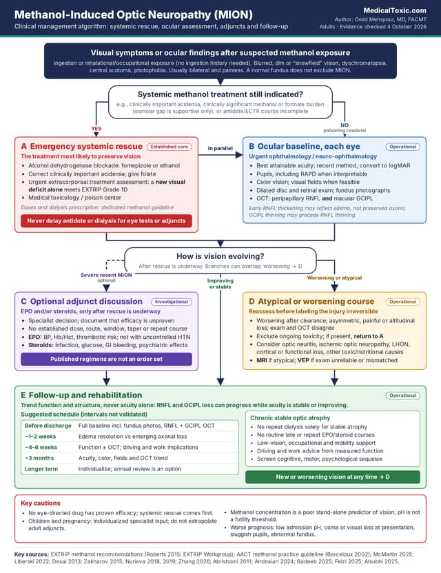
Methanol-induced optic neuropathy (MION) is primarily a consequence of formate-mediated mitochondrial injury. During active poisoning, the treatment most likely to preserve vision is rapid systemic treatment of the poisoning: alcohol dehydrogenase blockade, correction of clinically important acidemia, folate, and extracorporeal treatment when indicated. A new visual deficit attributable to methanol is itself an EXTRIP Grade 1D indication for extracorporeal treatment; practical indications, modality selection, and dialysis management are addressed in the companion Extracorporeal Treatment (Hemodialysis) in Poisoning — Clinical Guideline. No treatment specifically directed at methanol-induced optic neuropathy has established efficacy. Erythropoietin (EPO) and high-dose corticosteroids remain investigational and must never delay antidote therapy or extracorporeal treatment. [1–5]
This guideline addresses treatment, ophthalmic assessment, prognosis, and follow-up of MION. It is intentionally not a general methanol-poisoning protocol. Detailed fomepizole or ethanol dosing, bicarbonate protocols, extracorporeal treatment criteria, and systemic methanol-poisoning management are covered in Methanol Poisoning: Clinical Recognition, Triage, and Treatment. The systemic treatment is summarized here only to the extent needed to protect vision. The systemic treatment is summarized here only to the extent needed to protect vision. The main pathway is written for adults; children and pregnancy require individualized toxicology and ophthalmology input. [1–5]
Evidence labels used in this guideline
Established emergency care: accepted toxicology management or an authoritative recommendation.
Investigational adjunct: plausible therapy supported mainly by case reports, case series, nonrandomized comparisons, or unpublished trial data.
Operational guidance: practical examination or follow-up steps that have not been validated as universal thresholds.
Clinical warning
A new visual deficit attributable to methanol is an EXTRIP Grade 1D indication for extracorporeal treatment. Alcohol dehydrogenase blockade, correction of clinically important acidemia, folate, and extracorporeal treatment when indicated take priority over ocular-directed therapy. [2–5]
EPO and high-dose corticosteroids have no established efficacy in MION. They are investigational and must never delay antidote therapy, metabolic stabilization, or extracorporeal treatment. [11–22]
Published EPO and corticosteroid regimens are not standard order sets; no preferred dose, route, treatment window, combination, repeat course, or steroid taper is established. [11–22]
Chronic stable optic atrophy after biochemical recovery does not by itself indicate repeat dialysis. EXTRIP recommendations apply to active or severe poisoning, including new visual deficits, not stable established optic atrophy. [3,4]
Clinical Issue | Recommendation |
|---|---|
Ocular baseline | As an operational baseline, document each eye separately: best attainable acuity, pupils, color vision, visual fields when feasible, optic disc/retina, and OCT of peripapillary RNFL plus macular ganglion-cell/GCIPL layers. Photograph the fundus when practical. [1,6–10] |
EPO and corticosteroids | Neither has established efficacy. Consider only case by case after systemic rescue is underway, preferably with neuro-ophthalmology and medical toxicology input and explicit discussion of uncertainty and harms. [11–22] |
Published regimens | May inform specialist discussion but are not standard order sets. No preferred dose, route, combination, treatment window, repeat course, or steroid taper has been established. [11–22] |
Follow-up | Do not use visual acuity alone. Structural loss can progress despite stable or improving acuity. Trend acuity, pupils, color, fields, RNFL, and ganglion-cell measurements over time. [7–10,23–25] |
Chronic stable optic atrophy | Does not by itself indicate repeat dialysis. EXTRIP indications apply to active or severe poisoning, not stable established optic atrophy. There is no evidence that late repeated EPO/steroid courses reverse established atrophy. Focus on surveillance, rehabilitation, and reassessment if vision worsens. [3,4,7,8,20–24] |
Clinical Phase | Priority | Action |
|---|---|---|
Active poisoning: new or worsening visual deficit | Emergency systemic rescue | Continue or commence alcohol dehydrogenase blockade; correct clinically important acidemia; give folate; urgently assess for extracorporeal treatment. New visual deficit alone meets an EXTRIP indication. Eye evaluation proceeds in parallel. [2–5] |
Recent MION after systemic rescue has begun | Define severity and document baseline | Urgent ophthalmology/neuro-ophthalmology assessment. Record function and structure. EPO and/or corticosteroids may be discussed only as investigational adjuncts; no validated treatment window or patient-selection rule has been established. [6,11–22,26] |
Biochemical recovery with persistent or evolving visual loss | Reassess before labeling irreversible injury | Confirm that ongoing systemic toxicity has been excluded; reconsider alternative diagnoses or evolving optic-nerve/retinal injury. Repeat OCT or MRI when it is expected to answer a defined clinical question; this is operational guidance rather than a validated testing threshold. [1,7,9,10,27] |
Chronic stable optic atrophy | Surveillance and rehabilitation | No evidence supports repeat dialysis or repeated ocular-directed drug courses solely for established atrophy; reassess EXTRIP indications if active toxicity or a new/worsening deficit is suspected. Continue functional/structural follow-up and low-vision support. [3,4,7–10,20–25] |
Methanol is metabolized to formate. Formate inhibits mitochondrial cytochrome-c oxidase and impairs oxidative phosphorylation. The retrolaminar optic nerve and retinal ganglion-cell system are especially vulnerable.
In the classic human histopathologic series, circumscribed myelin damage occurred immediately behind the lamina cribrosa with axonal preservation; later clinical and OCT literature also documents retinal ganglion-cell and axonal loss. Acute ATP failure can produce axoplasmic stasis and disc edema.
Later, RNFL/GCIPL thinning and optic atrophy may progress even after central acuity improves. [1,5,8–10,23,28]
MION is usually bilateral and painless. Symptoms may include:
Blurred or dim vision
Dyschromatopsia
Central or cecocentral scotoma
Photophobia
A “snowfield” or “snowstorm” visual description
The optic discs may appear normal early, become hyperemic or edematous, and later develop pallor or atrophy. A normal fundus does not exclude MION. [1,6,9]
Do not require a history of ingestion. Clinically important visual injury has been described after inhalational methanol exposure, including workplace exposures, so ask about relevant solvent or occupational exposure when the ingestion history is absent or implausible. [29]
A four-group visual-outcome classification reported by Sanaei-Zadeh et al. and later reproduced by Nagendran et al. describes:
Group 1: blurred or snowfield vision with complete recovery within up to 2 weeks after discharge.
Group 2: blindness at discharge with partial recovery within 3–4 weeks.
Group 3: blindness at discharge without subsequent improvement.
Group 4: blindness at discharge followed by partial recovery over days to about 1 month and later recurrent visual decline or blindness within up to 9 months.
Use this classification only to describe observed natural history, not to select treatment. [26,30]
Operational guidance: no universal minimum examination set has been validated. The following baseline is practical guidance synthesized from published MION cohorts and contemporary ophthalmic assessment. [1,6–10]
Best attainable visual acuity in each eye. Record the method and convert to logMAR when possible for serial comparison.
Pupillary reactions, including a relative afferent pupillary defect when interpretable.
Color vision when acuity permits.
Confrontation fields or formal perimetry when the patient can cooperate.
Dilated examination of the optic discs and retina.
Fundus photography when practical.
OCT of both peripapillary RNFL and macular ganglion-cell/GCIPL layers.
Early RNFL thickening may reflect edema; later thinning may reflect edema resolution, axonal loss, or both. [7–10]
Visual evoked potentials can detect persistent or subclinical visual-pathway dysfunction.
In a cohort of 47 methanol-poisoning survivors examined 1–9 months after the event, VEPs were abnormal in 20 patients (43%); five of those patients had normal Snellen visual acuity. [25]
Operationally, consider VEP when:
The clinical examination is unreliable.
Acuity and structural findings do not match.
Identification of persistent or subclinical pathway dysfunction would change counseling or follow-up.
MRI is not required to diagnose MION but may be useful when:
Deficits are asymmetric or atypical.
Neurologic findings are unexplained.
Another diagnosis is plausible.
Reported findings include optic-nerve diffusion restriction or enhancement and the systemic methanol pattern of bilateral putaminal injury.
A small retrospective series of 16 eyes found that visual acuity improved in eyes in which optic-nerve diffusion restriction regressed. This is a prognostic association, not a validated treatment-selection tool. [27]
Alternative Diagnosis | Features That Make Isolated MION Less Typical |
|---|---|
Other toxic/nutritional optic neuropathy | Gradual onset over weeks to months; relevant drug or nutritional history; usually no acute high-anion-gap metabolic toxicity |
Optic neuritis | Pain with eye movement, often unilateral/asymmetric, demyelinating features on MRI |
Ischemic optic neuropathy | Often unilateral, altitudinal field defect, disc-at-risk or vascular context |
Leber hereditary optic neuropathy | Sequential eye involvement, characteristic demographic/family context, no methanol metabolic syndrome |
Cortical visual loss | Preserved pupillary responses and normal optic discs; may coexist with methanol-related brain injury |
Functional visual loss | Inconsistent acuity or fields with normal pupils and no concordant structural/electrophysiologic abnormality; diagnose positively and cautiously after excluding organic disease |
During active methanol poisoning, emergency systemic rescue takes priority. Follow Methanol Poisoning: Clinical Recognition, Triage, and Treatment for ADH blockade, acid–base management, folate therapy, and extracorporeal treatment. Ocular-directed adjuncts must not delay antidote administration or extracorporeal treatment. [2–5]
The key clinical distinction is between:
A new or worsening visual deficit during active poisoning, and
Persistent stable optic atrophy after biochemical recovery.
Established optic atrophy alone is not an EXTRIP indication for repeated extracorporeal treatment. Worsening vision after apparent clearance should prompt urgent reassessment for ongoing toxicity, an alternative diagnosis, or evolving optic-nerve/retinal injury. [3,4]
Evidence status: Investigational
High-dose corticosteroids have been used with the rationale of reducing optic-disc or retrobulbar edema and inflammatory injury, but no adequately powered randomized trial has established improved visual outcome in MION.
The most frequently cited steroid-only evidence is a 2011 interventional case series of six patients. Visual acuity improved after:
Methylprednisolone 250 mg IV every 6 hours for 4 days
Followed by oral prednisolone 1 mg/kg/day for 10 days
Because the study had no comparator and the patients had already received systemic methanol treatment, spontaneous recovery and co-interventions cannot be separated from a steroid effect. [11]
Corticosteroids should not replace or delay:
Alcohol dehydrogenase blockade
Correction of metabolic toxicity
Extracorporeal treatment
If a specialist team elects to use corticosteroids for severe recent-onset MION, document that efficacy is unproven and that the regimen is extrapolated from uncontrolled literature.
No MION-specific evidence establishes a required oral taper after the short-course regimens reported in this literature. [11,13,14,18,21,22]
Assess:
Infection risk
Diabetes and hyperglycemia
Gastrointestinal bleeding risk
Psychiatric adverse effects
Hypertension
Relevant drug interactions
Monitor blood pressure, glucose, electrolytes, and mental status as clinically indicated. [31]
Evidence status: Investigational
EPO has neuroprotective and anti-apoptotic rationale, and several uncontrolled reports describe visual improvement. Human evidence remains very low certainty because severity, timing, dialysis and antidote treatment, steroid co-treatment, and spontaneous recovery differ substantially between reports. [12–22,32]
Study | Design | Main Finding | Why Certainty Remains Low |
|---|---|---|---|
Pakravan & Sanjari 2012 [12] | 2-patient case report | EPO 10,000 IU IV every 12 h for 3 days with high-dose methylprednisolone and vitamins/folate; marked recovery reported | No comparator; multiple simultaneous treatments |
Pakravan et al. 2016 [13] | 11 EPO + steroid vs 11 historical steroid controls | Both groups improved; final acuity favored EPO group | Nonrandomized historical control; small sample; baseline and temporal confounding |
Zamani et al. 2018 [14] | 10 EPO + steroid vs 5 steroid controls | Some early improvement in EPO-treated patients was followed by deterioration; no sustained advantage demonstrated | Very small case-control study; heterogeneous course |
Pakdel et al. 2018 [15] | 16 patients; prospective noncomparative series | EPO 20,000 IU IV daily for 3 days; visual acuity improved from baseline | No control group; some patients received repeat courses |
Pakdel 2019 / NCT02376881 [16,17] | Randomized, double-masked, placebo-controlled trial; results available only as conference abstract | Review-level reporting describes greater eye-level BCVA improvement with EPO | No peer-reviewed full report and no posted registry results identified; participant and eye denominators cannot be fully reconciled |
Tabatabaei et al. 2023 [18] | 105 treated patients; prospective uncontrolled cohort | EPO plus methylprednisolone associated with improvement from baseline | No untreated comparator; retreatment effect cannot be separated |
Alrobaian et al. 2024 [19] | 9 patients; retrospective comparison; 5 received EPO | Final visual acuity similar with and without EPO | Small study, but directly comparative and did not demonstrate a clear EPO advantage |
Badeeb et al. 2025 [20] | 8-patient retrospective series | No statistically significant pre/post acuity improvement; repeat EPO did not improve outcomes | Small heterogeneous series; argues against routine repeat courses |
Feizi 2025 and Alsubhi 2025 [21,22] | Two overlapping systematic reviews | Most reports describe improvement from baseline; comparisons with steroids alone remain conflicting | Primary evidence predominantly uncontrolled and co-treated |
EPO is not established routine therapy.
If a specialist team considers EPO for severe recent MION, treatment should occur only after emergency methanol management is underway, ideally within a research protocol or after explicit discussion that:
Efficacy remains unproven.
Optimal timing is unknown.
Optimal dose and route are unknown.
A preferred combination with corticosteroids has not been established.
Repeat treatment has not been shown to improve outcomes.
The strongest signal favoring EPO comes from a randomized trial available only in abstract form. The most informative published comparative cohort found similar final visual outcomes with and without EPO. [16,17,19,21,22]
Timing is unresolved.
Across the 2025 systematic-review literature, EPO was started approximately 2–29 days after visual symptom onset. These observational data do not define a therapeutic window and do not prove that earlier or later treatment is effective or ineffective. [22]
Epoetin products carry risks including:
Hypertension
Thrombosis
Stroke
Other cardiovascular events
Uncontrolled hypertension is a labeled contraindication.
Before use, review blood pressure, hemoglobin/hematocrit, thrombotic risk, formulation, and other contraindications with pharmacy. Small MION series are too small to establish safety. [33]
These regimens are descriptive and must not be converted into standard order sets.
Source | Published Regimen | Interpretation |
|---|---|---|
Abrishami 2011 [11] | Methylprednisolone 250 mg IV every 6 h for 4 days, then oral prednisolone 1 mg/kg/day for 10 days | Six-patient uncontrolled series |
Pakravan & Sanjari 2012 [12] | EPO 10,000 IU IV every 12 h for 3 days + methylprednisolone 500 mg IV every 12 h for 5 days, then prednisolone 1 mg/kg/day for 2 weeks | Two-patient report with multiple co-interventions |
Pakravan et al. 2016 [13] | EPO 10,000 IU IV every 12 h for 3 days with high-dose methylprednisolone | Nonrandomized comparison with historical controls |
Pakdel et al. 2018 [15] | EPO 20,000 IU IV once daily for 3 days; some patients received additional courses according to response | No control group; repeat-course benefit unproven |
Pakdel 2019 trial abstract [16,17] | EPO 20,000 IU IV in 100 mL normal saline over 2 h for 3 consecutive days vs placebo | Randomized trial, but results are only abstract-level and no registry results are posted |
Tabatabaei et al. 2023 [18] | EPO 10,000 IU IV every 12 h for 3 days + methylprednisolone 250 mg IV every 6 h for 3 days; monthly single-dose retreatment during follow-up | No untreated or steroid-only comparator for the main cohort |
Alrobaian et al. 2024 [19] | EPO 20,000 IU IV daily for 3 consecutive days, administered as daily infusion over 45 min | Retrospective 9-patient comparison; final visual outcomes were similar with and without EPO |
There is no evidence-based preferred:
EPO dose
EPO route
Corticosteroid regimen
Steroid taper
EPO/steroid combination
Retreatment schedule
Treatment window
Do not prescribe automatic monthly EPO pulses, prolonged steroid tapers, or repeated combined courses solely because they appeared in uncontrolled reports. [11–22]
Proposed ocular therapies beyond systemic rescue—including intravitreal EPO, vitamin-based regimens without documented deficiency, and other neuroregenerative approaches—are not established treatments for restoring vision after MION.
Correct documented nutritional deficiencies or electrolyte abnormalities on their own merits.
There is no established role for a prolonged folate course solely to reverse established optic neuropathy once systemic toxicity has resolved. [1,21,22,26,32]
Visual acuity alone is an incomplete outcome.
Long-term cohorts show that structural and electrophysiologic injury may persist or progress even when central visual acuity improves.
A 50-patient Czech follow-up cohort found long-term visual sequelae in approximately 40% of patients. In a 42-survivor prospective cohort, 31% had abnormal RNFL thickness and 24% had chronic axonal loss; progressive visual loss occurred in 7 of 10 patients with chronic axonal degeneration. A 2026 longitudinal study of 17 patients found progressive RNFL and GCIPL thinning even in some patients whose acuity remained normal. [7–9,23]
The following schedule is operational guidance. Exact intervals have not been validated.
Time | Suggested Operational Follow-Up |
|---|---|
During hospitalization | Repeat visual acuity, pupils, symptoms, and fundus examination when clinically meaningful. Repeat OCT only if it is expected to change assessment or management; there is no evidence-based requirement for OCT every 24–48 h. |
After stabilization / before discharge | Establish a reproducible baseline: acuity, pupils, color, fields when possible, fundus photography, RNFL and macular ganglion-cell/GCIPL OCT. |
~1–2 weeks | Early ophthalmology/neuro-ophthalmology review when practical; confirm trajectory and distinguish edema resolution from emerging axonal loss. |
~4–6 weeks | Repeat functional testing and OCT if clinically useful; review occupational, driving, and daily-function implications. |
~3 months | Reassess acuity, color, fields, and OCT trend. Persistent or progressive deficits warrant longer follow-up. |
Longer term | Individualize. For persistent deficits or structural progression, ongoing surveillance—including annual review as an operational option—is reasonable. Worsening vision at any time requires earlier reassessment. |
At each meaningful follow-up, compare trends rather than isolated measurements.
Early RNFL thickening can represent edema; a subsequent fall may reflect edema resolution, axonal loss, or both. Macular ganglion-cell/GCIPL thinning may become apparent before RNFL thinning.
Visual evoked potentials can remain abnormal despite recovered Snellen visual acuity. [8–10,23,25]
Predictor | Clinical Interpretation |
|---|---|
Admission pH | Among the most consistent prognostic markers. Lower pH is associated with worse visual outcomes. In the 4-year RNFL cohort, pH <7.3 was associated with chronic global RNFL loss. Do not use a pH threshold as a futility criterion. [6,8] |
Visual deficit or coma at presentation | Associated with persistent visual sequelae in long-term cohorts, but severe early visual loss can still partially recover. [7] |
Pupillary reactivity / fundus abnormality | Reduced pupillary reactivity and abnormal fundus findings correlate with worse outcomes in observational cohorts. [6] |
Delay to treatment | Longer delay has been associated with worse outcomes in some observational series, including inhalational poisoning, but findings are not uniform and confounding by severity is substantial. [6,29] |
Admission methanol concentration | Poor stand-alone prognostic marker because it measures parent compound rather than accumulated formate burden. Do not use it alone to predict vision. [1,5,6] |
ApoE4 carriage | Associated with abnormal visual-pathway function in one 4-year cohort. Research finding only; not a routine clinical test. [23] |
Optic-nerve diffusion restriction | Regression of diffusion restriction correlated with better acuity in a small retrospective MRI series. Exploratory, not a validated treatment-selection tool. [27] |
Counseling should remain cautious.
Visual recovery ranges from substantial improvement to permanent severe visual loss. Long-term follow-up studies also show that sequelae may be missed at hospital discharge.
In the six-year Estonian follow-up, 8 of 22 examined patients who had been discharged without recognized visual sequelae were later found to have visual disturbances, although the study had substantial loss to follow-up. [24]
For persistent impairment, consider:
Low-vision rehabilitation
Occupational support
Mobility support
Medication-management assistance
Counseling regarding driving
Counseling regarding safety-sensitive work based on measured function and applicable requirements
Assess cognitive, motor, and psychological sequelae after severe poisoning because neurologic injury may coexist.
Explain that visual improvement can occur, but complete recovery cannot be promised and apparent recovery of central acuity does not eliminate the need for follow-up. [7,8,23,24]
The principle that active methanol toxicity must be treated urgently to protect vision remains applicable, but systemic and ophthalmic management should be individualized with medical toxicology, pediatric, and ophthalmology input.
Do not automatically extrapolate investigational adult EPO or high-dose corticosteroid regimens to children. [2–5]
Urgent systemic treatment of active methanol poisoning remains the priority. Management should involve medical toxicology, obstetric, and ophthalmology input.
Evidence for ocular-directed EPO or high-dose corticosteroids in pregnancy is even more limited than in typical adult MION cohorts. Do not extrapolate an investigational adult regimen automatically. [2–5]
Intervention | Current Evidence | Clinical Position |
|---|---|---|
Systemic antidote + metabolic stabilization + extracorporeal treatment when indicated | Authoritative toxicology guidance; new visual deficit is an EXTRIP Grade 1D indication | Established emergency care; highest priority during active poisoning [2–5] |
High-dose corticosteroids | One small steroid-only case series plus frequent use as co-treatment in EPO studies; no adequately powered randomized trial | Investigational; not routine standard therapy [11,13,14,18] |
IV EPO | Case reports/series, small nonrandomized comparisons, one favorable randomized trial reported only as abstract, a small negative comparative cohort, and two overlapping systematic reviews | Investigational; case-by-case specialist decision [12–22] |
EPO + corticosteroid | Most published experience uses combination treatment, but major cohorts are uncontrolled and one case-control study did not show sustained benefit | No proven superiority over steroid alone, EPO alone, or natural recovery after detoxification [13,14,18,21,22] |
Repeat/prolonged EPO or steroid therapy | No comparative evidence; repeat EPO did not improve outcomes in a recent small series | Do not use routinely [15,18,20–22] |
Other ocular adjuncts | Evidence consists mainly of case-report, preclinical, or extrapolated data | Not routine therapy; research context only [21,22,26,32] |
The 2025 EPO systematic reviews consolidate the literature but do not convert EPO into established therapy. Their primary studies overlap substantially, most are uncontrolled, and comparisons with corticosteroids alone are conflicting. [21,22]
The 2024 Saudi comparative series and 2025 Badeeb series temper enthusiasm for routine EPO: one found similar final visual outcomes with and without EPO, and the other found no statistically significant pre/post improvement and no benefit from repeat courses. [19,20]
The 2026 OCT studies strengthen the case for longitudinal structural monitoring because RNFL and ganglion-cell loss can progress despite stable or improving acuity. [9,10]
No peer-reviewed full report or posted results were identified for NCT02376881 as of this update. ClinicalTrials.gov lists planned enrollment of 24 participants, while a 2022 review summarizes the conference abstract as an eye-level analysis of 34 eyes. Because the primary full report is unavailable, the favorable randomized-trial signal remains abstract-level evidence and the denominators should not be treated as fully reconciled. [16,17]
Publication of complete randomized EPO trial data and adequately powered randomized trials of EPO with and without corticosteroids.
Standardized outcomes that include visual acuity, visual fields, RNFL, macular ganglion-cell/GCIPL metrics, and patient-centered visual function rather than acuity alone.
A validated treatment window and clear definitions of acute, subacute, and chronic MION for interventional studies.
A validated surveillance schedule and prospective data linking OCT/VEP trajectories to functional outcomes.
Better correlation of visual injury with formate burden rather than parent methanol concentration.
The October 2026 MedicalToxic MION drafts were reconciled into a single focused treatment guideline. A targeted literature verification was performed through October 4, 2026 using PubMed-indexed studies, accessible full text, trial-registry information, EXTRIP recommendations, and current prescribing information, with emphasis on EPO, corticosteroids, OCT, visual outcomes, prognosis, and 2024–2026 publications.
Earlier pivotal histopathology and long-term cohort studies were retained where they directly inform current care.
This is a clinical evidence synthesis, not a de novo systematic review, independent dual-reviewer process, or new GRADE exercise. EXTRIP grades are retained.
Ocular-adjunct evidence is limited by small samples, heterogeneous severity and timing, simultaneous detoxification and dialysis, frequent EPO-steroid co-treatment, eye-level analyses, inconsistent outcome measures, and selective follow-up.
Improvement from baseline must therefore not be interpreted as proof that an ocular adjunct caused recovery.
Keep reading